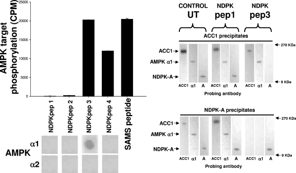
FIG. 2.
NDPK peptides. The left histogram shows pure, active rat liver AMPK used to phosphorylate a selection of peptides as follows. Four peptides (NDPKpep1, 2, 3 and 4) corresponding to regions of NDPK-A (amino acids 49 to 62, 92 to 105, 111 to 125 and 139 to 152, respectively) were used, and the SAMS peptide was used as a positive control, each at 1 mM concentration. For the left overlay, the overlay protocol involves a slight variation of the above-described Western blotting protocol in that each of the NDPK-A peptides was immobilized on a nitrocellulose membrane and then blocked/washed as described above for the Western blot protocol. Furthermore, 10 μg of pure, recombinant AMPK α1 or α2 protein was overlaid onto the peptides, with binding being detected using isoform-specific antibodies as described above for Western blotting. In the right blots, 10 μg of rat liver cytosol either left untreated (UT) or incubated with a molar excess of NDPKpep1 (pep1) or NDPKpep3 (pep3) was precipitated using an ACC1 (upper right) or NDPK-A (lower right) antibody and probed with either ACC1-, AMPK α1 (α1)-, or NDPK-A (A)-specific antibodies as labeled at the bottom of each lane. NDPKpep3 specifically interferes with the ACC1/AMPK α1/NDPK-A complex association.

