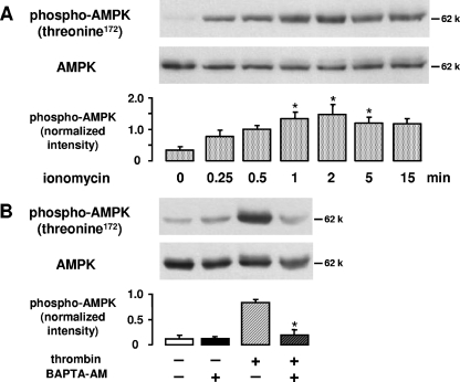
FIG. 3.
Role of Ca2+ in thrombin-induced AMPK activation. A. HUVEC were stimulated with ionomycin (2 μM) for the indicated times. B. HUVEC were pretreated with BAPTA-AM (20 μM, 30 min) and subsequently stimulated with thrombin (1 U/ml, 1 min). For panels A and B, the degree of threonine 172 phosphorylation of AMPK was determined by Western blot analysis with an anti-phosphospecific threonine 172 antibody. AMPK was stained with an antibody against AMPKα. Representative figures and densitometry data (means ± SEMs) of three independent experiments for each treatment are shown. Phosphospecific signals from ionomycin-stimulated and nonstimulated cells (A) or from cells stimulated with thrombin in the absence or presence of BAPTA-AM (B) were compared.  , P < 0.05.

