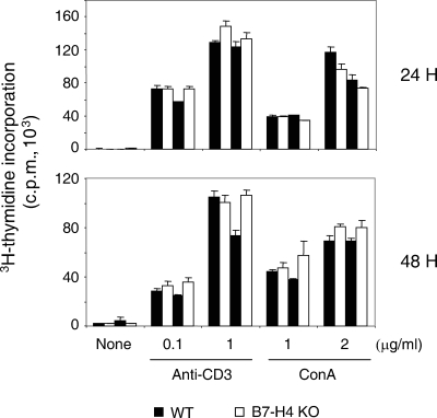
FIG. 2.
Normal T-cell proliferation in the absence of B7-H4. Single-cell suspensions of total LN cells were stimulated with soluble anti-CD3 (145-2C11; 0.1 or 1 μg/ml; BD Pharmingen) or concanavalin A (ConA; 1 or 2 μg/ml; Sigma) in U-bottom 96-well plates (1 × 10⁵ cells/well) in triplicate. The cells were pulsed with [3H]thymidine (1 μCi/well) for the last 8 h of a 24- or 48-h incubation period. The data shown are the mean ± the standard deviation from two WT and two B7-H4 KO mice and are representative of four independent experiments.

