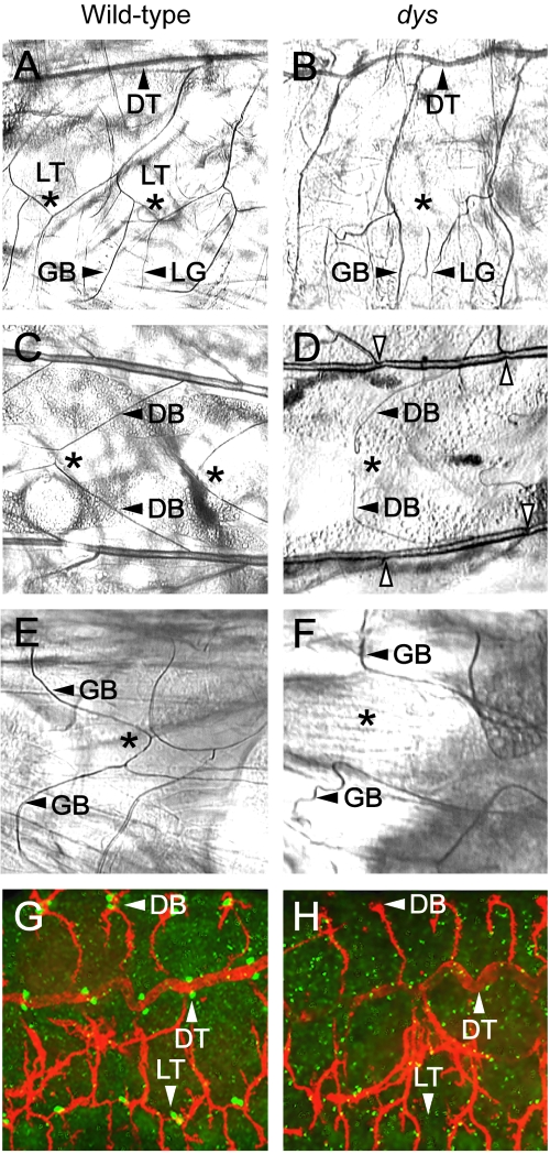
FIG. 2.
dys mutant embryos show tracheal fusion defects. Anterior is to the left in all panels. (A to F) Trachea from wild-type and dys2/dys3 mutant second-instar larvae were examined by phase microscopy. (A and B) Sagittal view. (A) The LT is fused (*) in a wild-type larva. The DT, GB, and lateral branch G (LG) are shown. (B) A dys mutant larva shows an absence of LT fusion (*). The GB and LG branch still persist and project ventrally. The DT is fused. (C and D) Dorsal view. (C) Wild-type larva showing fusion (*) of the DBs. (D) dys mutant larva showing a lack of DB fusion (*), although branches (arrowheads) are close together. Constrictions are seen in dys mutant DTs at fusion sites (open arrowheads). (E and F) Ventral view. (E) Wild-type larva showing fusion (*) of GBs. (F) dys mutant larva showing a lack of GB fusion (*). (G and H) Sagittal view. Confocal microscopic analysis of whole-mount embryos. (G) Stage 16 wild-type embryo stained with MAb 2A12 (red) and anti-Dys (green), showing fusion (arrowheads) of DBs, DTs, and LTs. Mab2A12 stains the tracheal lumen, and anti-Dys stains the tracheal fusion cells. (H) Stage 16 dys mutant embryo stained with MAb 2A12 (red) and anti-Dys (green), showing an absence of fusion of DB and LT branches. The DT is fused. Note the absence of anti-Dys reactivity.

