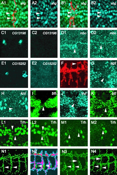
FIG. 6.
Regulation of gene expression in tracheal fusion cells. Each panel consists of a projection of three consecutive 1-μm confocal slices. (A to D) shg upregulation in DBs is dependent on dys function. Stage 16 shg-lacZ embryos were stained with anti-β-galactosidase (A1 and B2; blue), MAb 2A12 (A1 and B1; red), and anti-Dys (not shown). The use of anti-Dys staining allows for selection of homozygous mutant embryos, since they do not possess antibody-reactive protein. Fusion cells (arrowheads) express a higher level of β-galactosidase than surrounding tracheal cells in wild-type DB (A1 and A2). This upregulation was not observed in dys mutant DB fusion cells (B1 and B2, arrowheads). (C1 to M2) Embryos were hybridized to gene-specific probes and a dys RNA probe and immunostained with anti-Dys. dys RNA hybridization revealed fusion cells, and anti-Dys allowed identification of dys mutant embryos. (C1 and C2) CG13196 fusion cell expression is dependent on dys. Stage 15 wild-type DT fusion cells express CG13196 (C1), but dys mutants (C2) do not. (D1 and D2) mbo expression is regulated by dys. Fusion-4 embryos, which contain an mbo-lacZ enhancer trap gene, were immunostained with anti-β-galactosidase. Stage 15 wild-type DT (D1) fusion cells (arrowheads) show high levels of lacZ expression, whereas β-galactosidase expression is absent in dys mutant DT (D2). (E1 and E2) CG15252 fusion cell expression is dependent on dys. Stage 15 wild-type DT fusion cells (E1) express CG15252, but dys mutants (E2) do not. (F) Misexpression of CG13196 in btl-Gal4 UAS-CG13196 embryos results in ectopic tracheal branch fusion (arrowhead). (G to K) Stage 15 embryos were hybridized to apt (G), kni (H), trh (I), vvl (J), and btl (K) probes as well as to a dys probe to mark fusion cells (not shown). RNA levels of all five genes are downregulated in fusion cells but are not regulated by dys (not shown). (L1 to M2) dys downregulates Trh protein levels. Trh protein levels are reduced in wild-type (L1) DT fusion cells (arrowheads) and (M1) DB fusion cells (arrowheads). In dys mutant embryos, Trh proteins levels increased to levels comparable to those in other tracheal cells in both DTs (L2) and DBs (M2). (N1 to N4) Embryos stained with ant-Trh (green). (N1) w− control embryo. Trh is localized to nuclei at uniform levels throughout the trachea. Cells of the spiracular branch (arrowheads) show the same levels of Trh as other tracheal cells. (N2 and N3) btl-Gal4 UAS-dys embryo. (N2) Embryo stained with MAb 2A12 (red), anti-Dys (blue), and anti-Trh (green). btl-Gal4 UAS-dys is expressed at high levels in all tracheal cells except the spiracular branch cells (green; arrowheads). (N3) Trh levels are reduced in dys-positive cells (arrow) compared to spiracular branch cells (arrowheads). (N4) btl-Gal4 UAS-dys-Δb embryo. Trh levels are uniform throughout the trachea, as indicated by comparable Trh levels in dys-positive cells (arrow) compared to spiracular branch cells (arrowheads).

