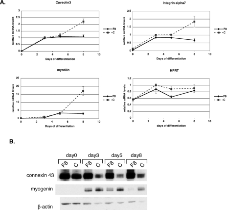
FIG. 3.
Myogenic differentiation defects in cells lacking FLNc. (A) Quantitative RT-PCR was used to measure the amount of various muscle transcripts throughout cell differentiation. On day 0, cells were switched to the low-serum condition. All amounts were normalized to an internal control (Tbp) and then to the amount of transcription in control cells at day 3. The experiment was performed in triplicate, and the error bars represent the standard errors. The three muscle genes shown here, caveolin3, myotilin, and α7 integrin, have a very low expression on day 0 both for control (C) and Flnc8 (F8) cell lines. The expressions of all three genes increases in both cell lines until day 5, after which the transcript level in the control line (dashed line) continues to increase, whereas the Flnc8 (solid line) does not show any change. Constitutively expressed Hprt does not show a significant difference in expression between the control and the Flnc knockdown line, indicating that the stall in transcription is specific to muscle genes. (B) Ten micrograms of total protein isolated from the C2C12 cell lines throughout myogenic differentiation was loaded onto 4 to 12% Bis-Tris gels (Invitrogen), and Western analysis was performed using connexin43- and myogenin-specific antibodies. Antibodies against β-actin were used to control for even loading.

