FIG. 6.
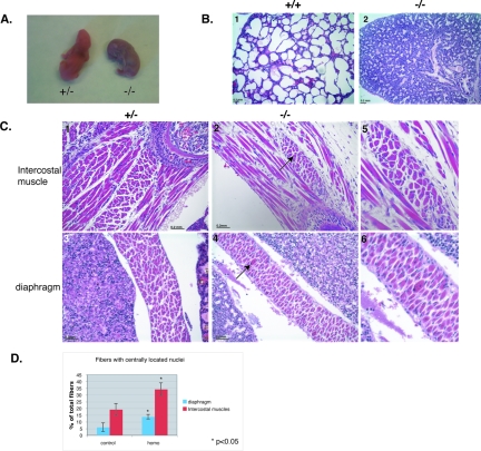
Flnc knockout mice die at birth, and muscles are myopathic. (A) All homozygous mice died within 10 min of birth and turned blue, whereas the control mice pinked up and continued breathing. (B) Hematoxylin (blue) and eosin (red) staining of the paraffin-embedded sections of mouse pups taken at ×10 magnification. The lungs of the homozygous mutants were not inflated (panel 2) compared to those of the wild-type littermates (panel 1). (C) Hematoxylin (blue, nuclei) and eosin (red, cytoplasm) staining of homozygous and control mice taken at ×20 magnification. The knockout mice have fewer muscle fibers and exhibit fibrosis. Muscles involved in respiration, such as intercostals, are severely decreased and show infiltration of connective tissue in homozygotes (panel 2) compared to the control littermates (panel 1). Other muscles, such as the diaphragm, show less drastic differences (panel 4 compared to panel 5) but still show centrally located nuclei (panel 5, arrow). Panels 3 and 6 show the magnified images of the centrally located nuclei in these muscles. (D) The percentage of the fibers with centrally located nuclei was counted in control animals (n = 2) and homozygous mutant animals (n = 3) in three different intercostal muscles and two random fields in the diaphragm. The percentage of fibers with centrally located nuclei was significantly increased in homozygous mutant mice (P < 0.05). The error bars represent the standard errors.

