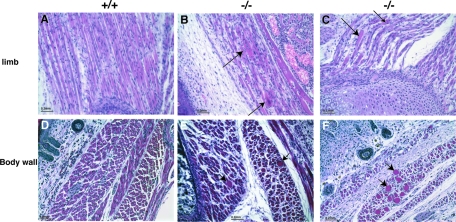
FIG. 8.
Abnormal muscle fiber pathology in Flnc knockout mice. Hematoxylin (blue, nuclei) and eosin (pink, cytoplasm) staining of muscles was visualized at ×20 magnification. Longitudinal sections show rounded fibers with centrally located nuclei (panels B and C, arrows). Cross-sections show vast variations in fiber size with giant fibers (panels E and F, arrows).

