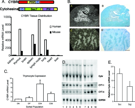
FIG. 1.
Cybr is expressed in hematopoietic tissues and regulated by cytokine stimulation. (A) Cybr expression was analyzed in RNA isolated from various mouse and human tissues by real-time PCR. (B) Dark field (a and c) and bright field (b and d) microphotographs of cryosections from adult mouse thymus (a and b) and spleen (c and d) were obtained after in situ hybridization with 35S-labeled antisense murine Cybr probes. Cybr expression is localized to the thymic medulla (a) and spleen white pulp (c). Counterstaining (b and d) was performed with 0.5% methyl green. (C) Mouse thymocyte populations were FACS sorted and analyzed for Cybr expression during thymic development by real-time PCR. Error bars indicate standard deviations. DN, double negative; DP, double positive. (D) Human peripheral blood mononuclear cells and MoDCs were stimulated with cytokine and analyzed for Cybr expression by Northern blotting. Peripheral blood mononuclear cells were cultured with media alone (lane 1) or stimulated with IL-2, IL-7, IL-12, IL-15, or IL-12 and IL-18 (lanes 2 to 6). Monocytes (lane 7), immature MoDCs (lane 8), or MoDCs matured with TNF-α treatment (lane 9) were also analyzed for Cybr expression. Unlike primary monocytes, the monocytic cell line THP-1 did not express Cybr (lane 10). (E) Cybr expression was analyzed in in vitro-polarized Th1 and Th2 cells by real-time PCR. Error bars indicate standard deviations.

