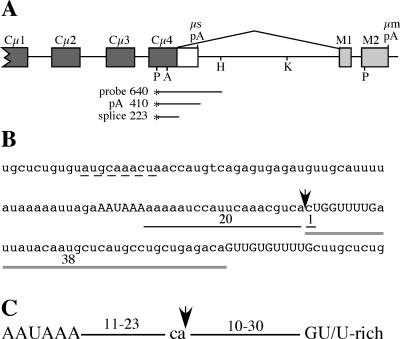
FIG. 1.
Structure of the immunoglobulin μ gene 3′ end, the μs poly(A) signal sequence, and poly(A) signal spacing. (A) The 3′ end of the Ig μ gene contains two cleavage-polyadenylation signals (μs pA and μm pA); the upstream μs poly(A) signal is in competition with the splice reaction between the Cμ4 and M1 exons (shown above the diagram). Filled boxes, constant-region exons; open box, μs-specific exon; light gray boxes, μm-specific exons. Restriction sites used in the cloning procedures are shown (P, PstI; A, ApaI; H, HindIII; K, KpnI). The S1 nuclease probe and the fragments protected by μs mRNA cleaved and polyadenylated at the μs poly(A) signal (pA) and μm mRNA that is spliced between Cμ4 and M1 exons (splice) are shown. (B) Sequence surrounding the μs poly(A) signal. The AAUAAA and two downstream GU/U-rich elements are shown in uppercase letters; the arrow indicates the cleavage site. The distances between AAUAAA and the cleavage site and the cleavage site and downstream elements are shown. The sequences suggested to match the consensus Oct1/2 binding site (9) and to contain a suboptimal U1A binding site (33) are underlined with a dashed line. (C) Standard spacing of the essential poly(A) signal sequence elements, as determined by comparison of poly(A) signal sequences and functional analyses as summarized in the text.

