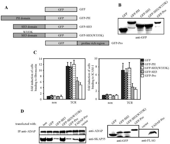
FIG. 4.
Isolated SH3 domain of SKAP55 or the proline-rich region of ADAP interferes with TCR-mediated activation of β1- and β2-integrins. (A) Scheme showing the GFP fusion proteins utilized in this study. (B) Jurkat T cells were transfected with either GFP, GFP-SKAP55-PH (GFP-PH), GFP-SKAP55-SH3 (GFP-SH3), GFP-SKAP55-SH3(W333K) [GFP-SH3(W333K)], or GFP-ADAP-Pro (GFP-Pro), and the expression of the GFP fusion proteins was assessed by anti-GFP Western blotting. (C) Transfectants were assessed for adhesion as described in the legend to Fig. 1C. non, nonstimulated; TCR, stimulated. Data represent the means and SE of at least three independent experiments. (D) Nontransfected (non) Jurkat cells or cells transfected with either GFP, GFP-SKAP55-SH3 (GFP-SH3), GFP-SKAP55-SH3(W333K) [GFP-SH3(W333K)], GFP-ADAP-Pro (GFP-Pro), FADAP-Pro, or empty vector (vector) were lysed, and endogenous ADAP was immunoprecipitated using a polyclonal sheep anti-ADAP serum. Precipitates (left) and total lysates (right) were analyzed by Western blotting for the presence of ADAP, SKAP55, GFP, or FLAG-tagged ADAP-Pro. IP, immunoprecipitation.

