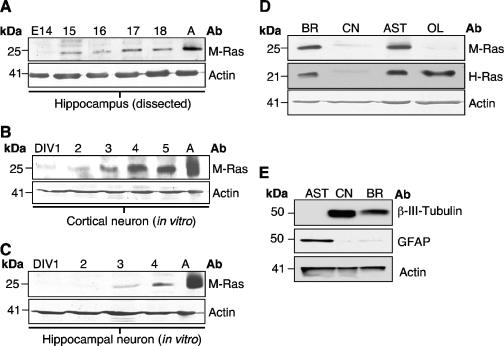
FIG. 2.
Expression of M-Ras in the CNS. (A) The expression of M-Ras in microdissected hippocampi from different embryonic stages was analyzed. Cell extracts from DIV cultures of cortical (B) and hippocampal (C) neurons were analyzed for M-Ras expression. Total cell extracts derived from adult brain (A) were used as a positive control. (D) A total brain lysate and primary cultures of cortical neurons (CN), astrocytes (AST), and oligodendrocytes (OL) were analyzed for the expression of both M-Ras and H-Ras. Note the lack of M-Ras expression in OL. (E) Lysates from DIV5 astrocytes, cortical neurons, and total brain extracts were subjected to Western blotting analysis with antibodies against GFAP and β-III-tubulin. All lysates were probed with an anti-actin antibody to ensure similar protein loading.

