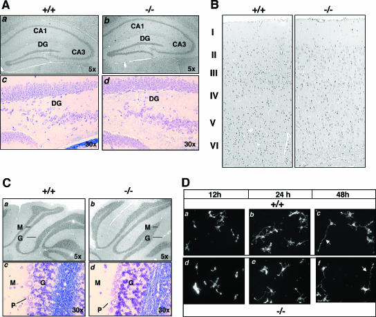
FIG. 4.
Brain morphology of Mras−/− mice. (A) Hippocampal regions of wild-type (+/+, panels a and c) and M-Ras KO mice (−/−, panels b and d) are compared. Hematoxylin-and-eosin (H&E) (panels a and b)- and Nissl (panel c and d)-stained paraffin sections show the CA1, CA3, and dentate gyrus (DG) regions, with the original magnifications indicated. (B) Coronal sections of cerebral cortex were stained with H&E, and the six-layered structure is indicated. (C) H&E (panels a and b)- and Nissl (panels c and d)-stained coronal sections of cerebellum from WT (+/+, panels a and c) and M-Ras KO (−/−, panels b and d) mice show the molecular (M), granular (G), and Purkinje (P) cell layers. (D) Primary hippocampal neurons were prepared from WT (+/+, panels a to c) and M-Ras KO (−/−, panels d to f) mice. At the indicated time points, cells fixed with 4% paraformaldehyde and actin were stained with Texas Red-conjugated phalloidin (converted to grayscale). Note the polarized extension of a single axon from the cell body (red arrows, panels c and f). Magnification, ×10.

