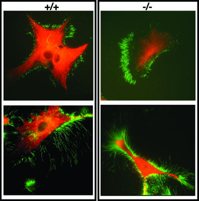
FIG. 5.
Morphology of M-Ras KO astrocytes. (A) Immunofluorescence analysis of WT (+/+) and M-Ras KO (−/−) astrocytes. Astrocytes are stained with a Cy3-conjugated anti-GFAP antibody (red), and the actin cytoskeleton at the cell periphery was detected with an anti-phospho-ERM antibody (green). Two representative astrocytes are shown for each genotype. Magnification, ×60.

