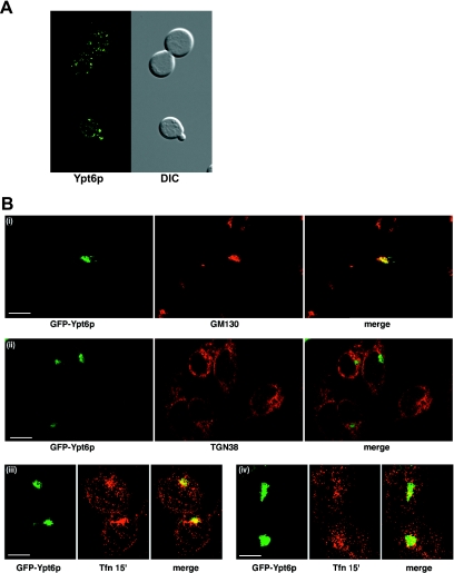
FIG. 3.
Localization of GFP-Ypt6p in yeast and HeLa cells. (A) GFP-Ypt6p was expressed in yeast cells, and live cells were viewed by epifluorescence microscopy. The GFP fluorescence signal is presented with differential interference contrast (DIC) images of the same cells. (B) GFP-Ypt6p was expressed in HeLa cells, and fixed cells were viewed by confocal microscopy. The GFP fluorescence signal is compared to those of antibody markers for the following cellular compartments: (i) Golgi apparatus GM130, (ii) trans-Golgi network TGN38, and (iii and iv) transferrin internalized for 15 min. In each case, a merge of the two fluorescence images is shown. Bars = 10 μm.

