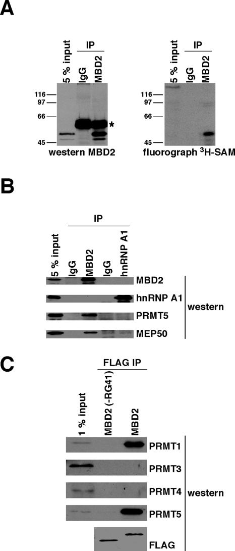
FIG. 4.
MBD2 coimmunopurifies with PMT and interacts with PRMT1 and PRMT5 in cells. IP, immunoprecipitation; IgG, immunoglobulin G. (A) Endogenous MBD2 complexes were immunoprecipitated from total human cell (Ramos) lysate by using MBD2 antibody, and IgG was used as a control. The immunoprecipitations were washed, resuspended in buffer containing [3H]SAM, and incubated at 37°C for 90 min. MBD2 was detached from the antibody beads by boiling in SDS sample buffer and analyzed by Western blotting with anti-MBD2 antibody or fluorography. Input represents cell lysate used for immunoprecipitations. The asterisk indicates the antibody heavy chain. (B) Endogenous MBD2 and hnRNP A1 complexes were immunoprecipitated from total human cell (Ramos) lysate. Corresponding IgGs were used as controls. The immunoprecipitations were boiled in SDS sample buffer and analyzed by Western blotting with the indicated antibodies. Input represents the total cell lysate used for the IP. (C) Human (293T) cells were transiently transfected with plasmids expressing FLAG-MBD2 or FLAG-MBD2(−RG41) proteins. FLAG proteins were prepared as described in the legend to Fig. 3B and analyzed by Western blotting with the indicated antibodies. Input represents pooled lysate from all transfections.

