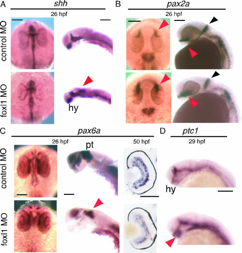
FIG. 4.
Expression pattern of shh and its related genes in foxl1 MO-injected embryos. Expression of shh, pax2a, pax6a, and ptc1 was examined by whole-mount in situ analysis. Embryos injected with control MO or foxl1 MO were harvested at the indicated stages, and whole-mount in situ hybridization was done. Expression patterns of shh, pax2a, pax6a (dorsal view with anterior at the top and lateral view with dorsal to the right), and ptc1 (lateral view with anterior to the left) are shown. Cross sections of retina are shown in the right panels of pax6a. Embryos with eyes removed are shown in the right panels of shh and the middle panels of pax6a. The results depicted were obtained with antisense probes. Sense probe control experiments were done for all of the probes, and no significant signals were detected (data not shown). The colored arrowheads are as defined in the text. Abbreviations: hy, hypothalamus; pt, pretectum. Scale bars, 100 μm.

