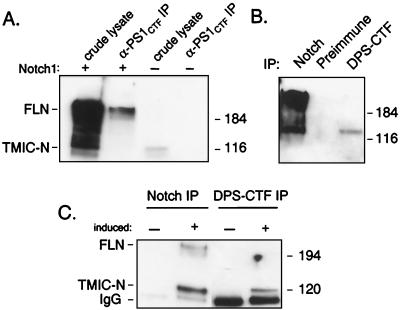
Figure 6.
Interaction between PS1 and Notch at endogenous expression levels. (A) 293 cells were transfected with completely full-length, wild-type mouse Notch1 bearing no epitope tag (+) or were untransfected (−) and either lysed in SDS lysis buffer and analyzed directly (crude lysate) or immunoprecipitated with α-PS1CTF. Following SDS/PAGE, Notch1 was detected with antibody mN1A. Crude lysates represent 20% of the protein analyzed in the immunoprecipitations (IP). (B) Drosophila clone 8 cells were lysed in coimmunoprecipitation lysis buffer and immunoprecipitated with the indicated antibody or with preimmune serum. Precipitating proteins were resolved by SDS/PAGE followed by Notch intracellular domain immunoblot. Each lane represents an equal amount of lysate. (C) A Drosophila S2 cells stable cell line expressing a metal-inducible promoter driving expression of Notch were either untreated (−) or induced with 700 μM copper sulfate overnight (+). Cells were then lysed in coimmunoprecipitation lysis buffer and immunoprecipitated with Notch or DPS-CTF as indicated. Precipitating proteins were analyzed by SDS/PAGE followed by Notch immunoblot. In this experiment, IgG heavy plus light chain was detected and is indicated. Sizes of molecular weight markers is given on the right.

