Figure 4.
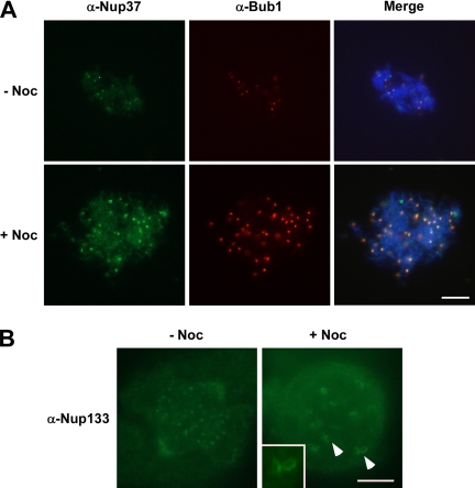
The Nup107-160 complex shows increased accumulation on unattached kinetochores. (A) Mitotic (CSF) extract was incubated with demembranated sperm chromatin for 40 min at 23°C with (bottom) or without (top) nocodazole. Mitotic chromosomes were collected and spread by centrifugation onto coverslips and then analyzed by indirect immunofluorescence with antibodies to Xenopus Nup37 (green) and Bub1 (red). The chromosomal DNA was stained with DAPI. Note that these conditions do not maintain spindle structure. Both Nup37 and BubR1 staining of kinetochores increases greatly. Bar, 10 μm. (B) HeLa cells were treated with nocodazole for 2 h before performing immunofluorescence by using affinity-purified anti-hNup133 antisera. The amount of the Nup107-160 complex, visualized with affinity-purified anti-hNup133 antisera, increased greatly giving large crescents, and ring structures typical of those outer kinetochore proteins that are affected by microtubule disassembly. Bar, 5 μm.

