Figure 6.
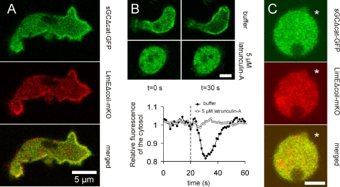
Colocalization sGC and filamentous actin. (A) sGCΔcat-GFP was coexpressed with the filamentous actin binding protein LimEΔcoil-mKO in gc-null cells. The cells were placed in a cAMP gradient and allowed to migrate toward the cAMP. Localization of sGCΔcat-GFP is shown in the top panel, LimEΔcoil-mKO in the middle panel, and the overlay in the bottom panel. The arrow points in the direction of migration. (B) gc-null cells expressing the N-terminal localization domain of sGC fused to GFP (N-sGC1-1019) were globally stimulated with 10−6 M cAMP at t = 20 s, as indicated by the dashed line. Translocation of the localization domain to the cell cortex was monitored in the absence (open circles) and presence of 5 μM latrunculin A (solid circles) by measuring the depletion of the cytosolic fluorescence. Bar, 5 μm. (C) gc-null cells coexpressing N-sGC1-1019 and LimEΔcoil-mKO were treated with 5 μM latrunculin A. A pipette filled with 10−4 M cAMP was placed next to the cells, at the position indicated by the mark. The localization of N-sGC1-1019 is shown in the top panel, LimEΔcoil-mKO in the middle panel, and the overlay in the bottom panel. Bar, 5 μm.

