Figure 1.
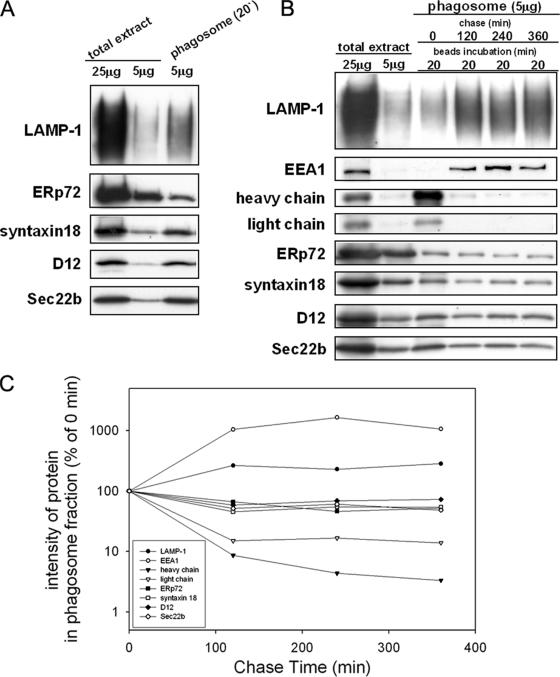
ER-localized SNARE proteins are enriched in isolated phagosomes. (A) J774 macrophages were incubated in the presence of nonopsonized latex beads for 20 min, after which the phagosome fraction was isolated from the cells by sucrose density centrifugation as described in Materials and Methods. The total cell extract (25 μg and 5 μg) and the isolated phagosome fraction (5 μg) were analyzed by SDS-PAGE and subsequent Western blotting with the use of antibodies against LAMP-1 (a lysosome marker), ERp72 (an ER marker), and ER-localized SNARE proteins (syntaxin 18, D12, and Sec22b). (B) J774 cells were incubated in the presence of IgG-opsonized latex beads for 20 min and then washed to remove the unbound beads. After further incubation for the indicated time (120, 240, or 360 min), the phagosome fraction was isolated from the cells by sucrose density centrifugation. The total cell extract (25 and 5 μg) and the isolated phagosome fraction (5 μg) were analyzed by SDS-PAGE and subsequent Western blotting with various antibodies as indicated. EEA1 is an early endosome marker protein. The heavy and light chains were derived from the IgG used for opsonization of the beads. (C) The signal intensity on the Western blot (B) was quantified by densitometry using NIH ImageJ (developed at the U.S. National Institutes of Health) and expressed as a percentage of the signal intensity at time 0.

