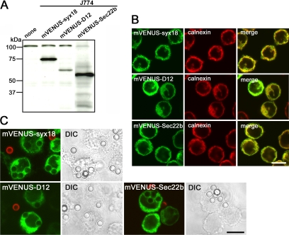
Figure 2.
mVENUS-SNARE proteins are recruited to phagosomes in J774 macrophages. (A) Western blotting analysis of total lysates from J774 cells stably expressing mVENUS-SNARE proteins. (B) J774 cells expressing mVENUS-SNARE proteins were fixed and stained with an antibody against an ER marker, calnexin. (C) J774 cells expressing mVENUS-SNARE proteins were incubated in the presence of IgG-opsonized latex beads for 20 min and then incompletely ingested beads were stained with Alexa 594–labeled anti-rabbit IgG antibodies (red) before fixation. Differential interference contrast (DIC) images indicate the location of the beads. Bars, 10 μm.

