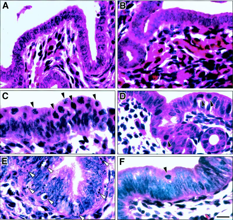
Figure 4.
Mitosis and apoptosis in wt (A, C, and E) and Igf1-null (B, D, and F) uterine epithelium. Mice were treated with vehicle (A and B) or E2 for 21 h (C and D) or 48 h (E and F). Solid arrowheads indicate some mitotic figures, and open arrowheads show examples of apoptotic bodies. The significantly greater occurrence of apoptosis in wt uteri shown here by hematoxylin/eosin staining was confirmed by analysis of DAPI-stained sections and by in situ end-labeling of fragmented DNA. (Bar = 10 μm.)

