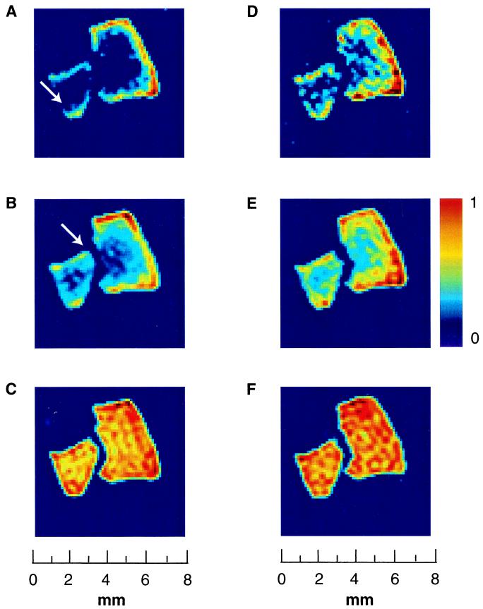
Figure 2.
Penetration of laser-polarized xenon into silica aerogel fragments at two different pressures as a function of time delay between excitation pulses. Shown are 2D magnetic resonance image slices perpendicular to the flow from a 3D data set, with a slice thickness of 100 μm and an in-plane resolution of 250 × 250 μm. (A–C) Magnetic resonance images of xenon adsorbed in aerogel fragments at high pressure (3 atm of Xe and 1 atm of N2; spectrum shown in Fig. 1A, dashed line around 25 ppm). The diffusion coefficient of xenon at T = 290 K under these conditions is Daero = 0.35 mm2/s. The pulse delay times are 0.2 s (A), 0.4 s (B), and 2 s (C). (D–F) Magnetic resonance images of xenon adsorbed in aerogel fragments at lower pressure (0.5 atm of Xe and 0.5 atm of N2; spectrum shown in Fig. 1A, solid line at 25 ppm). The diffusion coefficient of xenon at T = 290 K under these conditions is Daero = 0.65 mm2/s. The pulse delay times are 0.2 s (D), 0.4 s (E), and 2 s (F). The asymmetrical distribution of xenon spin density inside the aerogel fragments, for the shorter time delays, reflects the close proximity of fragments to each other and to the walls of the container, which attenuates the efficient accessibility of polarized gas to the fragments (white arrow).

