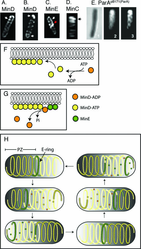
FIG. 6.
Helical organization of the MinD/ParA cytoskeletal proteins. (A to D) Fluorescently labeled MinD (A and B), MinE (C), and MinC (D). (Reprinted from reference 187 with permission of the publisher. Copyright 2003 National Academy of Sciences, U.S.A.) (A and B) Helical distribution of MinD when coexpressed with MinE. (A) A bright MinD helical structure is present at one end of the cell, representing a MinD polar zone (white arrow). A less intense coiled structure extends to the other end of the cell, presumably representing the underlying pole-to-pole MinD helical structure. (B) Short MinD polar zones are visible at both ends of the cell (white arrows), one presumably representing a growing zone and the other representing a polar zone at the opposite end of the cell, in the last stage of disassembly, as depicted in panel H. (C) Helical distribution of MinE when coexpressed with MinD. The majority of the MinE molecules are present in an end loop (the MinE ring, red arrow) of the MinE helical array that represents the polar zone. The white arrow indicates polar zone coils. Micrographs in panels A to C are deconvolved images. (D) Helical distribution of MinC when coexpressed with MinD and MinE (three-dimensional reconstruction from an optically sectioned cell). The arrow indicates the MinC coils of the polar zone. (E) Helical distribution of fluorescently labeled ParA from plasmid pB171. 1, Nomarski image; 2, raw image; 3, deconvolved image. (Reprinted from reference 39 with permission from Blackwell Publishing.) (F) Mechanism of MinD polymer growth on the cytoplasmic face of the inner membrane by addition of MinD-ATP subunits. (G) Stimulation of disassembly of MinD-ATP polymer by a MinE ring. MinE molecules in the E-ring stimulate the ATPase activity of the terminal MinD-ATP subunit in the polymer. Hydrolysis converts MinD-ATP to MinD-ADP, which is released from the end of the polymer. (H) Cyclic assembly and disassembly of the MinD helical polar zones (PZ) and MinE ring, leading to pole-to-pole oscillations. MinD-ATP subunits are yellow, MinD-ADP subunits are orange, and MinE subunits are green. See text for details. The solid yellow helical structures represent the underlying MinD helical structure that extends between the two ends of the cell. MinD helical structures also contain MinE and MinC, which are not shown for simplification.

