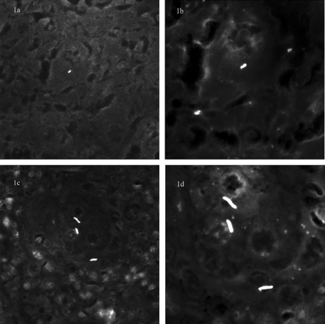
FIG. 1.
Comparison of morphometric perception of M. tuberculosis and M. avium subsp. paratuberculosis under 40× and 100× objectives. Auramine-rhodamine-stained sections of spleen from M. avium paratuberculosis- and M. tuberculosis-infected mice. (a) Under a 40× objective, providing ×400 total magnification, individual M. avium paratuberculosis forms are difficult to distinguish from artifacts. (b) Under an oil immersion objective, with total magnification ×1,000, individual M. avium paratuberculosis organisms are detectable. (c) In contrast, individual M. tuberculosis bacilli can be seen using a 40× objective and are further resolved using oil immersion (d).

