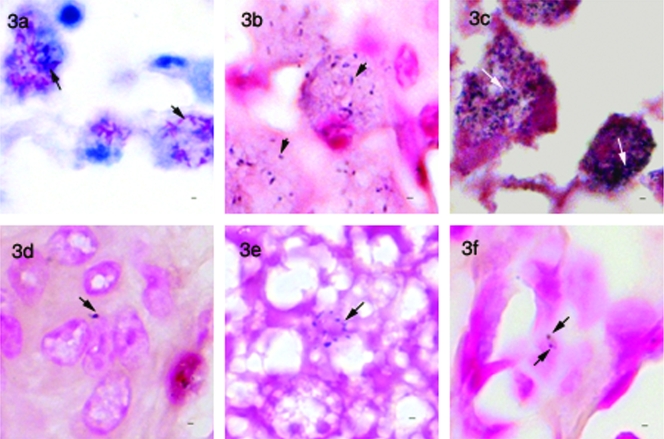
FIG. 3.
IS900-based in situ staining assays. (a) Ziehl-Neelsen-stained tissue section of ovine intestinal tissue showing a large number of mycobacteria in macrophages (arrows). (b) IS900-probe-based in situ hybridization of a sequential section of the same tissue, showing positive labeling (arrow) but a reduced number of signals compared to the Ziehl-Neelsen-stained section. (c) Indirect in situ PCR for IS900 of the same sample showing granular signals (arrows) and sensitivity comparable to that with Ziehl-Neelsen staining. (d) Section of liver from an M. avium paratuberculosis-infected mouse showing positive signal (arrow) by IS900-probe-based in situ hybridization. (e) Section of liver from an uninfected mouse and (f) section of lung from a mouse infected with M. tuberculosis showing nonspecific signals with the IS900-probe-based in situ hybridization method. Bar, 1 μm; magnification, ×950.

