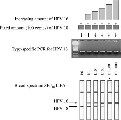
FIG. 1.
Competition between HPV genotypes. The top part is a graphical representation of the mixtures of the HPV-16 and HPV-18 plasmids prepared in this experiment. The middle part shows the agarose gel electrophoresis results of the type-specific HPV-18 PCR. The bottom part shows the results of SPF10 LiPA. The positions of the probes for types 16 and 18 are indicated (note that only the top part of the LiPA strips which contains the relevant probes is shown). The strip marked with as asterisk (1:1,000) showed a very weak reactivity with the HPV-18 probe line, which is not visible on the scans.

