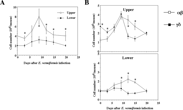
FIG. 3.
Cell number of IEL following infection with E. vermiformis. (A) Total IEL number was counted by staining with trypan blue. (B) Population of IEL bearing TCRαβ and TCRγδ of the upper and lower parts following infection. IEL were stained with fluorescein isothiocyanate-conjugated TCRγδ MAb, PE-conjugated TCRβ MAb, and allophycocyanin-conjugated CD3ɛ MAb. The TCRαβ and TCRγδ expression was gated on CD3+ cells. Three mice were used in each point. Values represent the mean ± standard deviation of three individual experiments in a triplicate assay. Asterisks represent statistically significant differences (P < 0.05).

