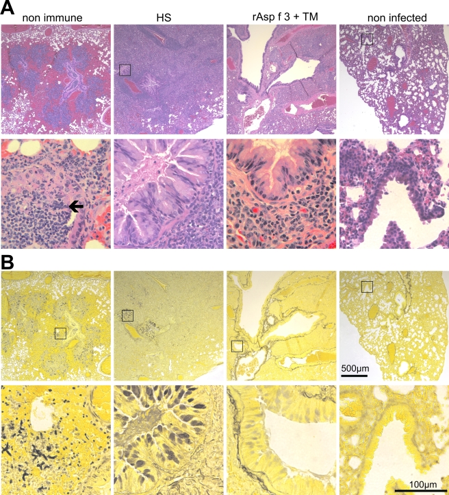
FIG. 6.
Histopathology. Hematoxylin and eosin (A) and Gomori silver (B) staining of consecutive slices of formalin-fixed lungs of a fatally infected nonimmune animal (first column), an HS-vaccinated survivor (second column), an rAsp f 3- plus TM-vaccinated survivor (third column), and a noninfected mouse (far right column). Magnifications, ×20 for the top row and ×200 for the bottom row in each panel. Square insets, when displayed in the top rows, mark the regions chosen for the higher-magnification image shown in the bottom row. The black arrow points to a hyphal structure that is readily visible in the hematoxylin and eosin-stained tissue.

