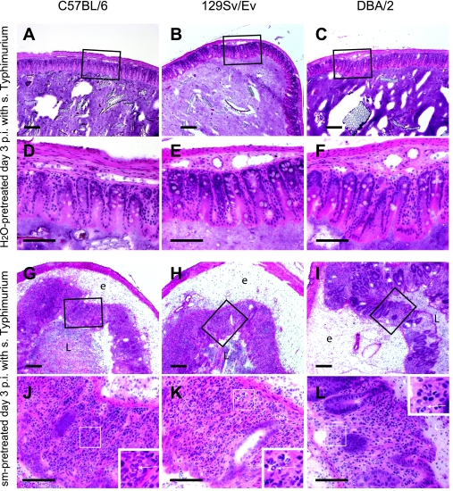
FIG. 2.
Pathological changes in ceca of H2O-pretreated or streptomycin-pretreated C57BL/6, 129Sv/Ev, and DBA/2 mice infected with wild-type serovar Typhimurium. Thin sections (thickness, 5 μm) of cecal tissues of mice from the experiment described in the legend to Fig. 1 were HE stained as described in Materials and Methods. (A to F) H2O-pretreated serovar Typhimurium-infected C57BL/6, 129Sv/Ev, or DBA/2 mice; (G to L) streptomycin-pretreated serovar Typhimurium-infected C57BL/6, 129Sv/Ev, or DBA/2 mice. e, edema; L, cecal lumen. Panels D to F and J to L (bars, 100 μm) are enlargements of the boxed areas in panels A to C and G to I (bars, 200 μm), respectively.

