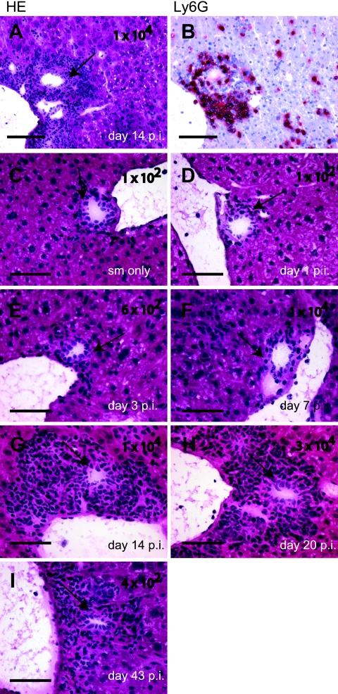
FIG. 7.
(A and B) Cholangitis in streptomycin-pretreated, serovar Typhimurium-infected 129Sv/Ev mice. Serial thin sections (thickness, 5 μm) of liver tissues of a 129Sv/Ev mouse infected for 14 days with serovar Typhimurium from the experiment described in the legend to Fig. 3 were HE stained (A) and immunohistochemically stained for Ly6G (B) as described in Materials and Methods. (C to I) Time course of the development of cholangitis. Streptomycin-pretreated 129Sv/Ev mice were from the experiments described in the legends to Fig. 1 and 3. Animals either were not infected (C) or were infected with serovar Typhimurium for 1 day (D), 3 days (E), 7 days (F), 14 days (G), 20 days (H), or 43 days (I). Bars, 200 μm (A and B) and 50 μm (C to I). Arrows point at gall duct epithelium; numbers are CFU/liver in the respective animal.

