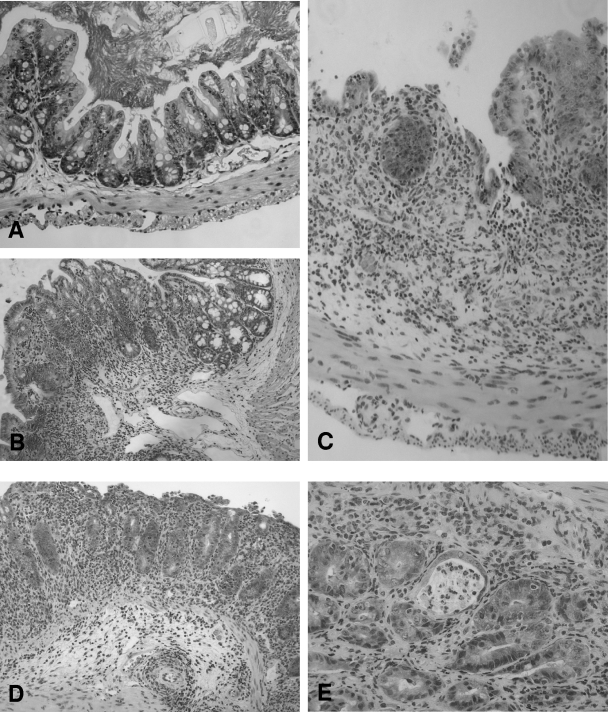
FIG. 4.
Histopathology of large intestine tissue from LF and LF-SCID mice 28 days postinoculation. (A) Only mild inflammation was detected in the lamina propria of LF mice colonized by C. jejuni, with preservation of the normal tissue architecture. (B to E) Severe inflammation was evident in the mucosa and submucosa of the cecum and colon tissue of similarly colonized SCID mice, with marked inflammatory infiltrate, including ulceration (B), epithelial hyperplasia and loss of goblet cells (C), and edema and architectural distortion (D). (E) Cryptitis was also frequently appreciated. Magnification, ×200.

