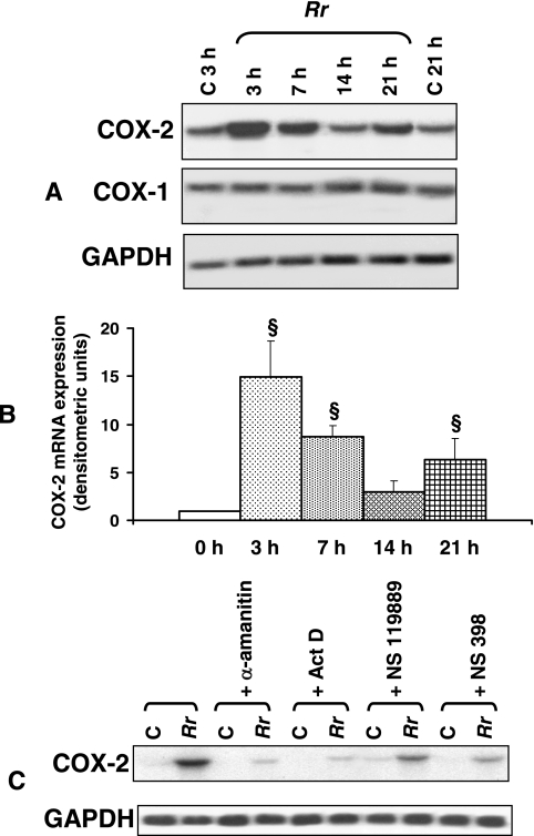
FIG. 1.
Time course of R. rickettsii-induced COX-2 expression in endothelial cells. (A) Northern analysis of RNAs isolated from uninfected EC at 3 and 21 h (C, control) and from cells infected with R. rickettsii (Rr) for 3, 7, 14, and 21 h. Blots were probed in succession with 32P-labeled human-specific COX-2, COX-1, and GAPDH (housekeeping gene) cDNA probes, and the results of a typical representative experiment are shown. (B) The steady-state levels of COX-2 mRNA in R. rickettsii-infected EC at different times postinfection were normalized to that of GAPDH and compared with the average values of basal levels in simultaneously cultured cells that were left uninfected. The mean baseline COX-2 expression level in each experiment was assigned a value of 1. The results are presented as means ± standard errors for a minimum of three independent experiments, and statistically significant changes from uninfected controls (0 h) are indicated by the symbol §. (C) Effects of the transcriptional inhibitors α-amanitin (5 μg/ml) and actinomycin D (Act D; 0.25 μg/ml), the protein synthesis blocker NSC 119889 (20 μM), and the selective COX-2 inhibitor NS 398 (50 μM) on R. rickettsii-induced COX-2 mRNA expression. C, uninfected controls; Rr, R. rickettsii-infected EC.

