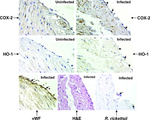
FIG. 5.
Immunohistochemical detection of COX-2 and HO-1 in R. rickettsii-infected endothelia from intact human umbilical veins. The serial sections of uninfected controls and infected cord specimens were stained with COX-2- and HO-1-specific antibodies. Sections from infected cords were stained for vWF and rickettsial antigen (indicated by large arrows) as described in Materials and Methods. Counterstaining with hematoxylin and eosin (H&E) was also carried out. Positive COX-2 (dark brown) and HO-1 (light brown) staining was predominantly detected in the endothelial cells of infected vasculature and is indicated by small arrows. Positive COX-2 staining in some inflammatory and mesenchymal cells of infected cord specimens was also seen. Magnification, ×400.

