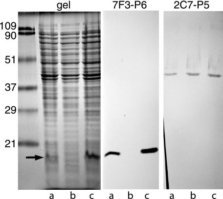
FIG. 1.
Coomassie blue-stained SDS gel (left panel) and immunoblot assays of whole bacterial cell lysates (middle and right panels). Lanes a, parent strain; lanes b, P6 mutant; lanes c, complemented P6 mutant. The center panel was probed with the monoclonal antibody 7F3, which recognizes an epitope on outer membrane protein P6. The right panel was probed with the monoclonal antibody 2C7, which recognizes an epitope on outer membrane protein P5. The arrow denotes the location of P6. Molecular mass markers are noted on the left, in kilodaltons.

