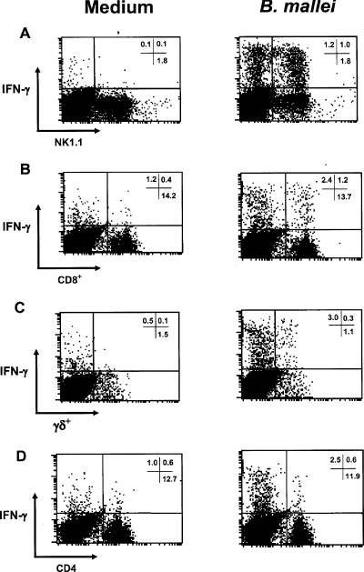
FIG. 7.
Cellular sources of IFN-γ following in vitro stimulation with heat-killed B. mallei. Splenocytes from uninfected C57BL/6 mice were incubated with medium alone or with 1 × 10⁶ heat-killed B. mallei cells/ml for 24 h prior to analysis by flow cytometry. The plots show intracellular IFN-γ in NK1.1+ cells (A), CD8+ T cells (B), TCRγδ T cells (C), and CD4+ T cells (D). The numbers in quadrants are the percentages of positive gated cells, and the data shown are representative of the data from three independent experiments.

