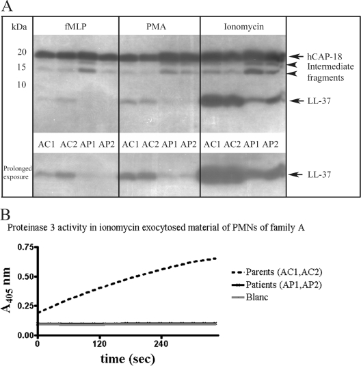
FIG. 2.
Immunoblot of hCAP-18/LL-37 of exocytosed material of PMNs isolated from PLS patients and their parents from family A. (A) Equal volumes (10 μl) of exocytosed material were analyzed. PMNs were stimulated with PMA (4 μg/ml), fMLP (2.5 μM), or ionomycin (1 μM) for 15 min at 37°C. Supernatants were analyzed by Western blotting with monoclonal antibodies to LL-37. hCAP-18 and LL-37 are indicated by arrows. Intermediate fragments indicate partially processed hCAP-18. The lower panel is the lower half of the immunoblot shown in the upper panel with a longer exposure time. Parents are indicates by AC1 and AC2; patients are indicated by AP1 and AP2. All samples were run on one gel. (B) Proteinase 3 activity in ionomycin exocytosed material of PMNs of family A. Samples (10 μl) were analyzed for proteinase 3 activity. Activity is expressed as the increase in absorption at 405 nm over time. Blanc, sample consisting of the reaction mixture was included in this assay to correct for spontaneous decay of the substrate. Note that the lines of the patients and the blanc coincide. The family A parents are indicated by AC1 and AC2; the family A patients are indicated by AP1 and AP2.

