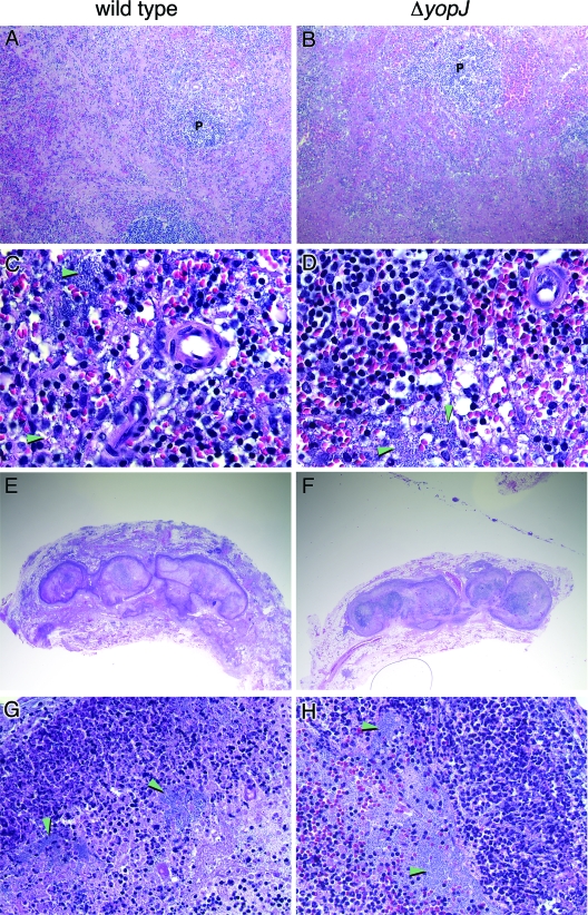
FIG. 2.
Identical histopathologies produced by wild-type and ΔyopJ Y. pestis. Hematoxylin-and-eosin-stained sections of spleens (A to D) and inguinal buboes (E to H) from rats infected with wild-type (left column) or ΔyopJ (right column) Y. pestis were examined. The arrowheads indicate large aggregates of extracellular bacteria. P, periarterial lymphatic sheath. Original magnifications, ×200 (A and B), ×600 (C and D), ×10 (E and F), and ×400 (G and H).

