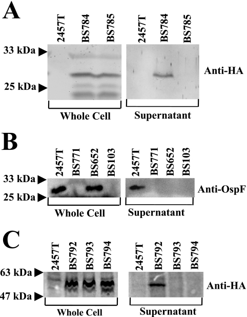
FIG. 1.
T3SS-dependent secretion of OspF and OspC1. Congo red was added to a final concentration of 7 μg/ml to activate the secretion of T3SS effectors. After 1 h, two sets of samples were saved: whole cell fractions and supernatant. OspF samples were run on a 12% SDS-PAGE gel, and OspC1 samples were run on a 10% SDS-PAGE gel. (A) Samples were immunoblotted with anti-HA antibody to visualize OspF-2HA (∼29-kDa band). (B) Samples were immunoblotted with anti-OspF antibody (∼27.5-kDa band). (C) Samples were immunoblotted with anti-HA antibody to visualize OspC1-2HA (∼54-kDa band).

