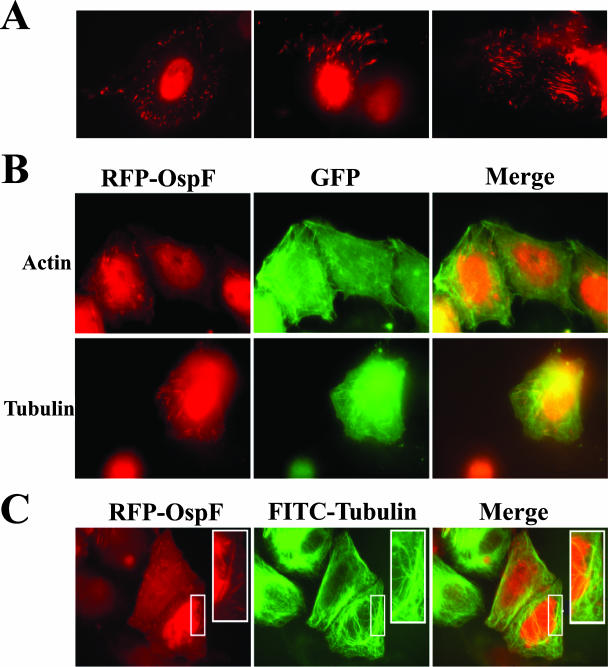
FIG. 6.
RFP-OspF localizes to a target in the cytoplasm. (A) HeLa cells were transfected with RFP-OspF. Each panel shows a different field of cells. (B) HeLa cells were cotransfected with RFP-OspF and GFP-actin or with RFP-OspF and GFP-tubulin. The panels on the left show the RFP-OspF signal (red), the panels in the middle show the GFP signal (green), and the panels on the right show the merged signals. (C) HeLa cells were transfected with RFP-OspF, fixed, and stained with anti-beta-tubulin conjugated to FITC. The panel on the left shows the RFP-OspF signal (red), the panel in the middle shows anti-beta-tubulin FITC signal (green), and the panel on the right shows the merged signals. The insets show magnified areas of interest. The micrographs are representative of the results of experiments repeated at least three times.

