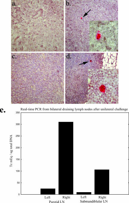
FIG. 4.
T. cruzi infection is limited to the ipsilateral side of challenge. (a to d) IHC staining of the right and left submandibular lymph nodes harvested from two separate mice 12 days after infection by contaminative challenge of the right eye. The left submandibular lymph nodes (a and c) show no signs of intracellular amastigotes, the replicative form of the parasite. In contrast, T. cruzi pseudocysts can be detected by immunohistochemistry in the right lymph nodes (b, b inset, d, and d inset). Proliferative centers were also seen in the right but not left lymph nodes (data not shown). (e) Further evidence for lateralization of infection by real-time PCR. Mice were infected by contaminative challenge of the right eye in the manner described above. The left eyes were exposed to an inactivated T. cruzi whole lysate prepared from numbers of parasites similar to those used for live challenge, and 10 days after infection the submandibular and parotid lymph nodes were harvested for DNA extractions. Real-time PCR was performed as described in Materials and Methods. Significant levels of parasites were seen in the right lymph nodes but not in the left.

