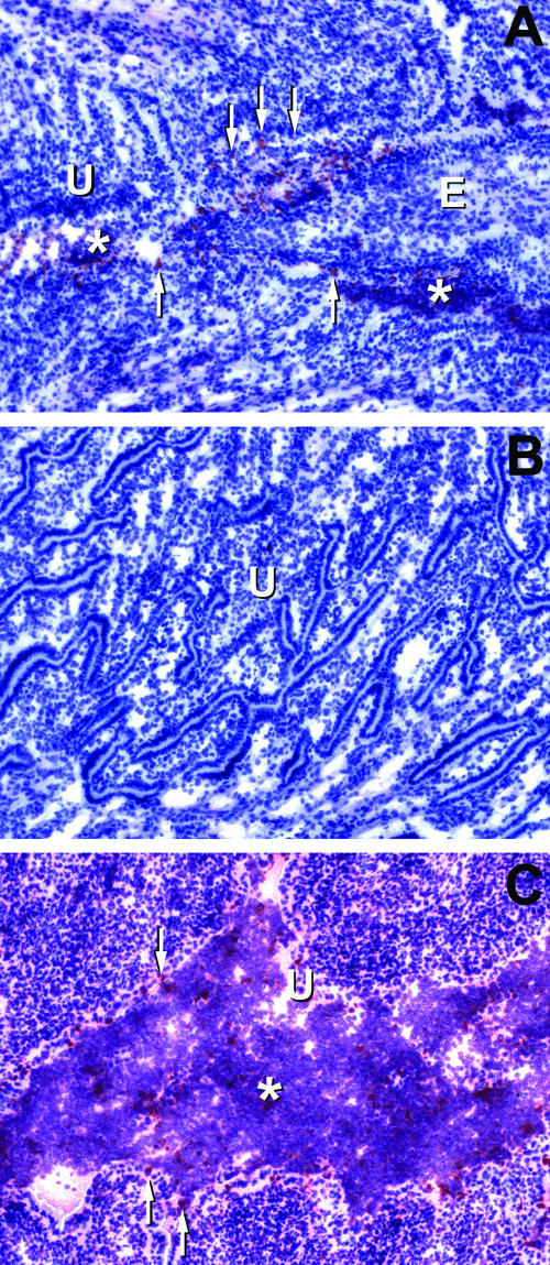
FIG. 6.
Immunohistochemical localization of chlamydial infection in mice treated with CMT-3. All histological sections shown are from day 7 postinfection. Chlamydial antigen staining appears red, whereas the counterstain with hematoxylin leaves the tissue pale blue. Panel A shows the juncture between the endocervix and the proximal uterus in a CMT-3-treated mouse. Diffuse chlamydial antigen staining is present within the lumenal structures, and condensed staining consistent with chlamydial inclusions is present in epithelial layers and scattered throughout the lumen. Panel B is the uterine horn endometrium of the same mouse as panel A. There was a lack of chlamydial antigen staining or inflammation in this region, which is atypical for this time point in the infection in normal, untreated animals. Panel C is the endometrium of the uterine horn of a vehicle-only-treated mouse and shows typical findings relative to chlamydial antigen staining, lumenal dilatation, and vigorous inflammatory infiltrates. Control slides of uninfected tissue or infected tissue treated with the secondary antibody only showed no chlamydial antigen staining. U, uterine endometrium; E, endocervix. Asterisks show areas of inflammatory cell infiltration, and arrows point to focal staining consistent with the localization and morphology of chlamydial inclusions in columnar epithelium.

