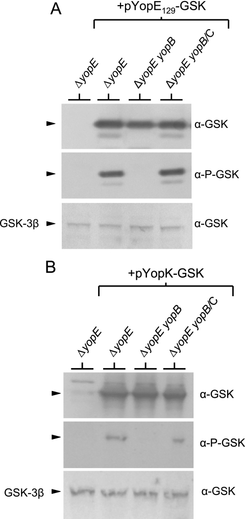
FIG. 3.
Injection and phosphorylation of YopE129-GSK and YopK-GSK require the expression of a functional YopB/YopD translocon. HeLa cell monolayers were infected at an MOI of 30 with Y. pestis KIM5-3001.P39 (ΔyopE) and KIM5-3001.P64 (ΔyopE ΔyopB) carrying plasmid pYopE129-GSK (A) or plasmid pYopK-GSK (B). Infected monolayers were subjected to SDS-PAGE and immunoblot analysis with anti-GSK antibodies (α-GSK) and phosphospecific anti-GSK antibodies (α-P-GSK). Levels of HeLa GSK-3β (used as a loading control) are shown. No translocation and phosphorylation of YopE129-GSK or YopK-GSK was detected in lysates from HeLa cell monolayers infected with the yopB deletion strain. Complementation (/C) of the yopB deletion strains with plasmid pYopB2 restored normal levels of YopE129-GSK or YopK-GSK translocation and phosphorylation.

