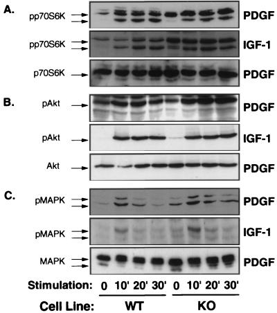
Figure 2.
PDGF- and IGF-1-stimulated activation of p70S6-kinase, Akt, and MAPK is unaltered in KO cells. Western blot analyses were performed on cell extracts from control (WT) and KO cells, which had been stimulated with PGDF or IGF-1 for the indicated times (min). Immunoblotting was performed with antibodies against phosphorylated p70S6-kinase (A, Top and Middle), phosphorylated Akt (B, Top and Middle), and phosphorylated MAPK (C, Top and Middle) or with corresponding antibodies, detecting these proteins independent of activation (A–C, Bottom)

