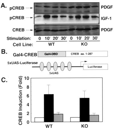
Figure 6.
PDGF- and IGF-1-stimulated phosphorylation and activation of CREB are unaltered in the absence of Rsk-2. (A) Western blot analysis using an anti-CREB antibody (Bottom) shows comparable cellular content of CREB in WT and KO cells. Western blot with an Ser-133 phospho-specific anti-CREB antibody reveals normal phosphorylation of CREB in KO cells in response to PDGF (Top) and IGF-1 stimulation (Middle). (B) Schematic representation of the heterologous CREB reporter system. (C) WT and KO cells were cotransfected with the described reporter plasmid. After serum depletion for 36 hr, cells were left untreated (open bars) or treated with PDGF (black bars) or IGF-1 (gray bars) for 4 hr before lysis. Data represent the stimulation of luciferase activity/β-galactosidase activity in stimulated vs. untreated cells.

