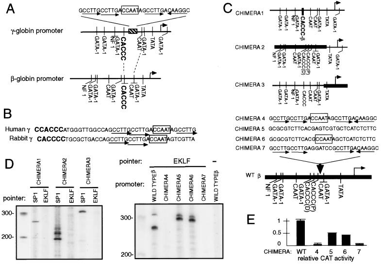
Figure 3.
Recruitment of EKLF to the γ-β chimeric promoters. (A) The human γ-globin promoter contains CCTTG repeats (striped box) in a region between the CACCC (bold letters) and the proximal CAAT (rectangle) boxes that is not present in the β-globin promoter. The position and orientation of each CCTTG repeat is marked with a horizontal arrow. The CACCC and CAAT boxes of the γ- and β-globin promoters are aligned with dashed lines. (B) The rabbit γ-globin promoter also contains the CCTTG repeat (horizontal arrow). (C) Diagram of γ-β chimeric promoters. In chimera 1, the CACCC box of the γ-globin promoter (thin line) was replaced with the CACCC box (CACCC-β) of the β-globin promoter. In chimera 2, the region upstream of the TATA box was replaced with the corresponding region of the β-globin promoter (thick line), and the minimal promoter region was from the γ-globin promoter (thin line). Chimera 3 is the opposite of chimera 2. In chimeras 4–7, the indicated sequences (31 bp) were inserted 3′ of the β-globin CACCC (proximal) box. (D) Recruitment of the EKLF pointer to the chimeric promoters in MEL cells. PIN*POINT assays were performed with EKLF pointer and a target plasmid containing the indicated chimeric promoter. (Left) The recruitment of Sp1 and EKLF pointer to chimeras 1 and 2 was detected by performing a primer extension with primer JS64 and the recruitment of Sp1 and EKLF pointer to chimera 3, with primer JS41. (Right) The recruitment of EKLF pointer to chimeras 4–7 was examined by performing a primer extension with primer JS41. The cleavage site in chimeras 5 and 6 were located ≈30 bp upstream of the cleavage site in the wild-type β-globin promoter because of the 31-bp insertion. (E) Suppression of EKLF pointer recruitment correlates with suppression of transcription. Chloramphenicol acetyltransferase (CAT) assays of the reporter constructs containing the γ-β chimera 4–7 were performed 48 hr after transiently transfecting them into MEL cells.

