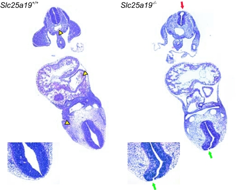
Fig. 3.
Coronal sections of unaffected (Left) and mutant (Right) E10.5 embryos. These images are higher magnifications of the last of the serial sections shown in Fig. 10 (154 and 130, respectively). Yellow arrowheads mark the erythrocytes in the heart and vessels in the section from the unaffected embryo. The rostral neural tube is not fused (green arrows) but is fused caudally (red arrow) in the mutant embryo.

