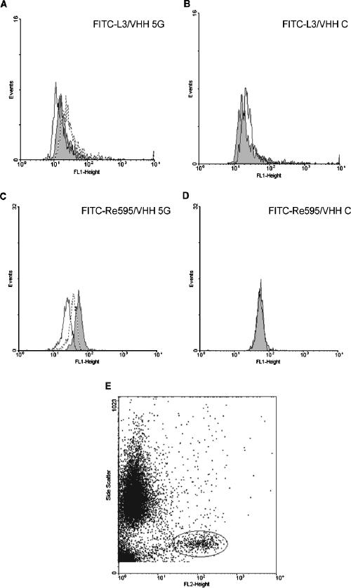
FIG. 4.
Inhibition of FITC-labeled LPS binding to monocytes. Fluorescence-activated cell sorting histograms showing the distribution of LPS-FITC associated with CD14-expressing cells. FITC-labeled L3 LPS from N. meningitidis (A) or FITC-labeled Re595 LPS from Salmonella serovar Minnesota (C) was incubated with isolated PBMCs in the absence of any VHHs (gray surfaces) or in the presence of 2 μg (stippled lines) or 10 μg (solid lines) of VHH 5G. The effect of an irrelevant VHH C (at 10 μg) on the distribution of the FITC-labeled L3 or Re595 LPS is depicted in panels B and D, respectively. Panel E shows the dot plot of PBMCs stained with the anti-CD14 MAb LeuM3-PE. Cells were gated via side scatter (y axis) and PE fluorescence.

