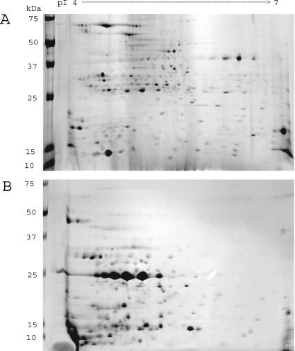
FIG. 3.
2-DE of M. paratuberculosis CE proteins (A) and CF proteins (B). Organisms were grown in WR medium and harvested at early stationary phase. After extraction for CE or concentration for CF, 100 μg of protein was applied to the first-dimension pH 4 to 7 nonlinear IPG strips. Second-dimension separation was done by SDS-PAGE on 10 to 20% acrylamide gels and visualized by brilliant blue R staining. The figure is representative of three independent experiments.

